The human TP53 gene organization as depicted over the past 20-plus years was simple: it produced a single transcript containing 11 exons encoding a single protein comprising 393 amino acids (Soussi and May 1989).
In 1996, Reisman et al. showed that intron 1 of the mouse and human TP53 gene encoded an untranslated transcript whose function is still unknown.
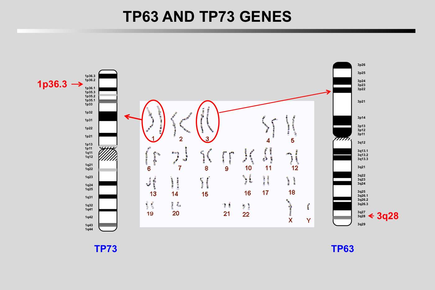
In 1997, Caput and coworkers revealed a first TP53 paralogs, TP73 localized on chromosome 1.
In 1997 and 1998, several groups revealed a second TP53 paralogs, TP63 localized on chromosome 3.
In 2005, Bourdon et al. identified two novel exons, beta and gamma, as well as a complex architecture involving various mechanisms to transcribe at least 8 mRNAs and translate up to 12 different protein isoforms. A second promoter localized in intron 4 lead to the expression of 4 TP53 transcripts encoding shorter TP53 isoforms.
In 2009, Mahmoudi et al. identified the WRAP53 gene that partially overlaps the 5' region of the TP53 gene in a head-to-head configuration. A similar gene is also found in the 5' region of the TP73 gene but it is lacking in the TP63 gene.
Click on the links below for further details
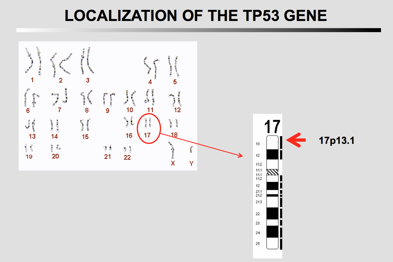
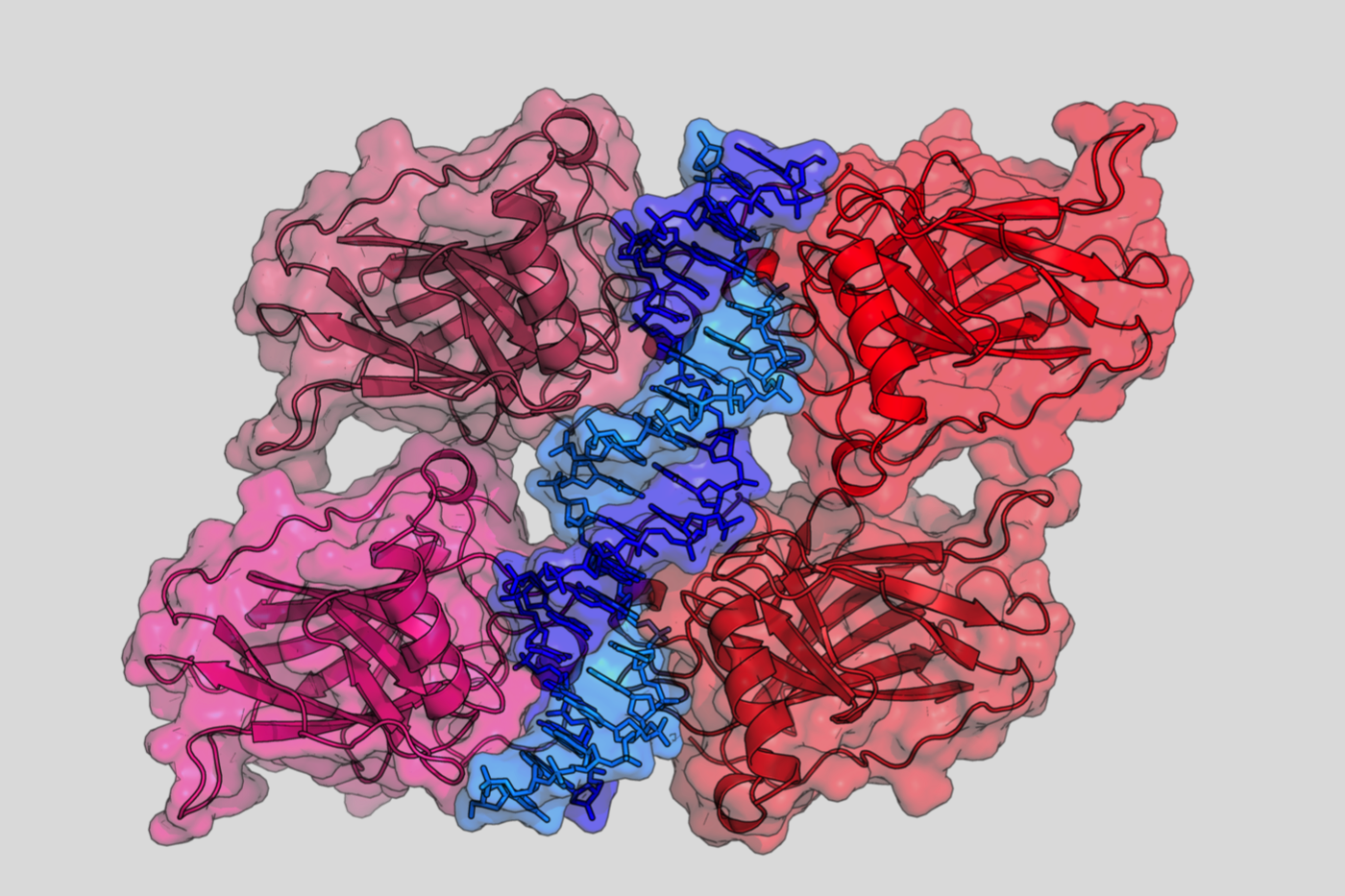
The p53 protein is encoded by the TP53 gene, which is highly conserved throughout evolution and is located on human chromosome 17p13.1.
LOH is frequently found on the chromosomal arms 17p in various types of human cancer.
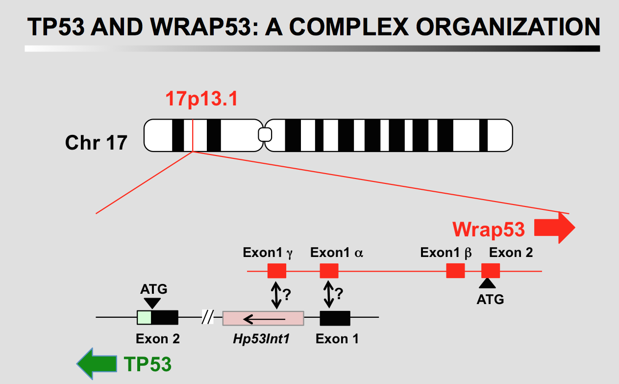
Organization of the 5' ends of the TP53 and WRAP53 genes. The two genes have a head-to-head configuration (Mahmoudi et al., 2009). The overlap between the transcripts (depicted by thin lines) would suggest that each gene could regulate the other via an antisense mechanism. Exon 1 of TP53 is not translated and Intron 1 is 10 kb long. Hp53Int1: untranslated transcript of unknown function.
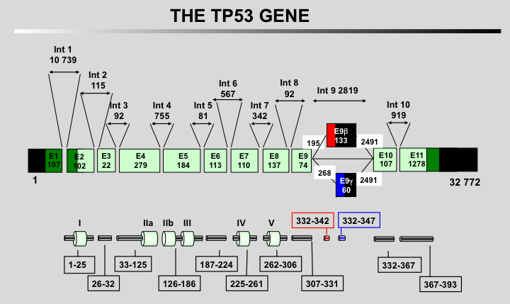
Structural organization for the human TP53 gene. Classical exons 1 to 11 are shown in green with exon 1 which is noncoding. Exons 2 to 11 are translated into a full-length TP53 protein (P1, 393 residues). Exons 9 beta (red) and gamma (blue) are used via an alternative splicing leading to different transcripts (see below).
Exons beta and gamma contain STOP codons and lead to truncated proteins missing part of the carboxy terminus of the protein. Translation of exons 9 beta and gamma adds 10 residues and 15 residues, respectively.
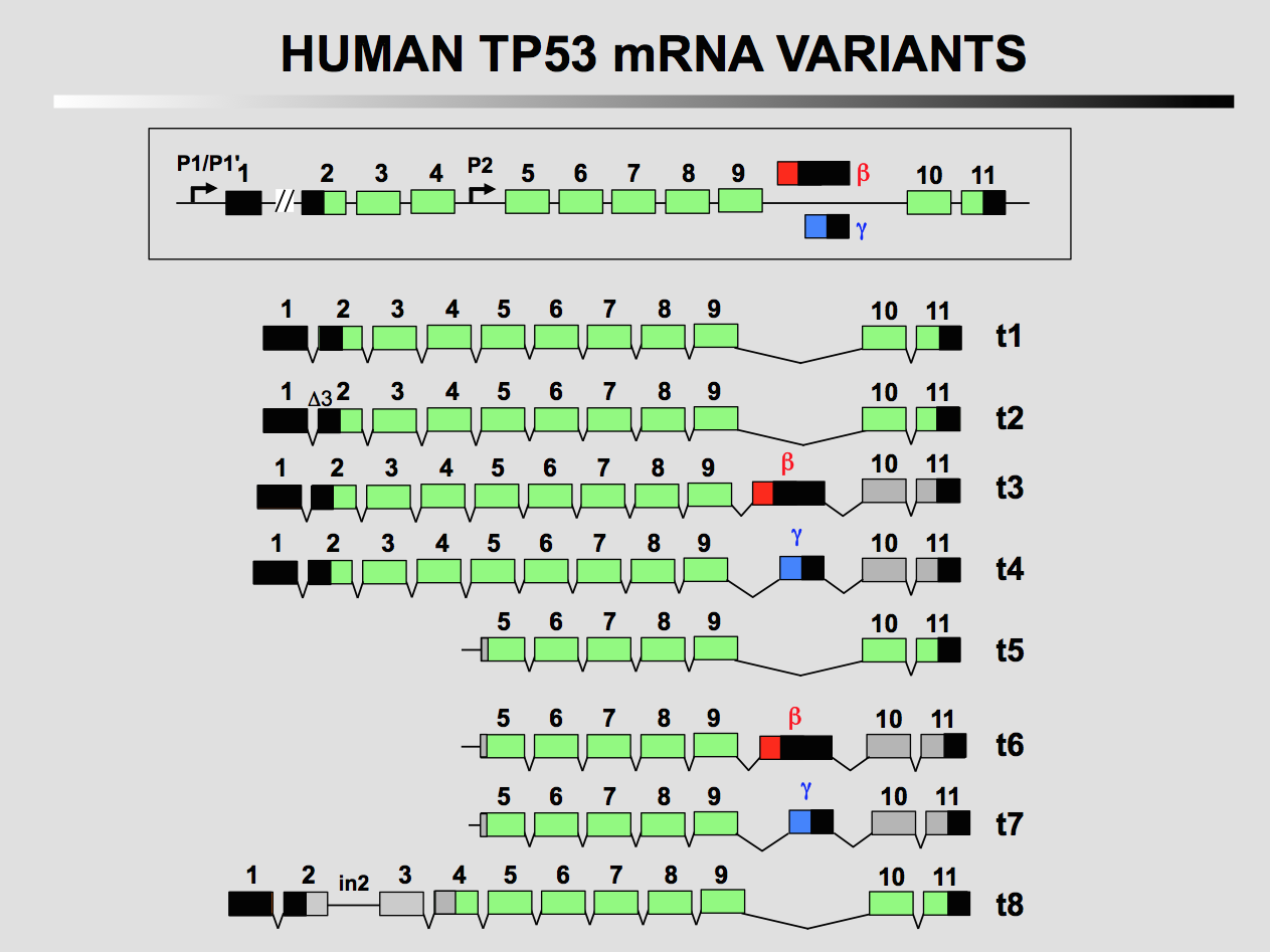
Transcriptional organization of the TP53 gene. The TP53 gene (upper part of the figure) is transcribed into 8 different mRNAs. Transcripts t1 to t4 originate from promoter P1 and P1' localized upstream to the gene. Transcripts t5 to t8 originate from promoter P2 localized in intron 4 and probably exon 3.
A full description of the various transcripts of the TP53 gene can be found at the LRG website
These links will open a new page
Reisman, D., Balint, E., Loging, W. T., Rotter, V., and Almon, E. (1996). A novel transcript encoded within the 10-kb first intron of the human p53 tumor suppressor gene (D17S2179E) is induced during differentiation of myeloid leukemia cells. Genomics 38, 364-370.
Soussi, T., and May, P. (1996). Structural aspects of the p53 protein in relation to gene evolution: a second look. J Mol Biol 260, 623-637.
Mahmoudi, S., Henriksson, S., Corcoran, M., Mendez-Vidal, C., Wiman, K. G., and Farnebo, M. (2009). Wrap53, a natural p53 antisense transcript required for p53 induction upon DNA damage. Mol Cell 33, 462-471.
Bourdon, J. C., Fernandes, K., Murray-Zmijewski, F., Liu, G., Diot, A., Xirodimas, D. P., Saville, M. K., and Lane, D. P. (2005). p53 isoforms can regulate p53 transcriptional activity. Genes Dev 19, 2122-2137.
Surget, S., Khoury, M. P., and Bourdon, J. C. (2013). Uncovering the role of p53 splice variants in human malignancy: a clinical perspective. Onco Targets Ther 7, 57-68.